Home

compozit Gară mineral cytoid bodies Zdrobi Adolescent Mare

Erythema, Purpura,Urticaria, Mastocytosis, EM, SJS and TEN - ppt video  online download
Erythema, Purpura,Urticaria, Mastocytosis, EM, SJS and TEN - ppt video online download

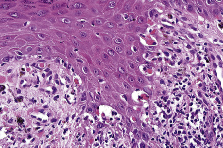
Moderate Melanin Incontinence and Some Evidence of a Late Lichenoid... |  Download Scientific Diagram
Moderate Melanin Incontinence and Some Evidence of a Late Lichenoid... | Download Scientific Diagram

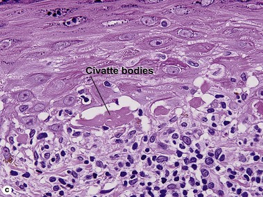
Civatte body in spinous layer in OLR {PAS after diastase treatment (X40)} |  Download Scientific Diagram
Civatte body in spinous layer in OLR {PAS after diastase treatment (X40)} | Download Scientific Diagram

Lichen planus ppt
Lichen planus ppt

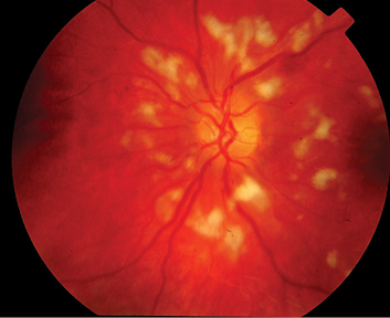
Cotton-Wool Spots May Challenge Diagnosis

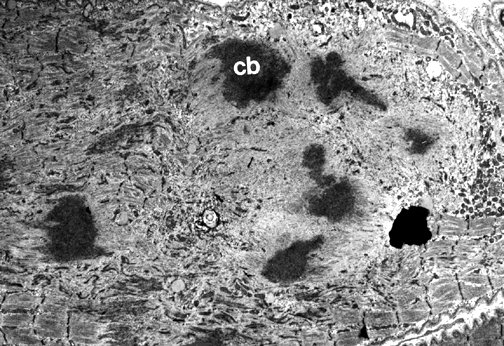
Light and electron microscopic studies on colloid and other cytoid bodies<link  href='#fn1'>*</link>
Light and electron microscopic studies on colloid and other cytoid bodies<link href='#fn1'>*</link>

Lichenoid and interface dermatitis | Basicmedical Key

Interface dermatitis | Plastic Surgery Key

Direct Immunofluorescence in Lichen Planus and Lichen Planus like Lesions -  World Journal of Pathology
Direct Immunofluorescence in Lichen Planus and Lichen Planus like Lesions - World Journal of Pathology

Lichenoid and interface dermatitis | Basicmedical Key

Cotton-Wool Spots (Inner Retinal Ischemic Spots) | Ento Key

Lymphohistiocytic infiltrate with occasional eosinophils in the... |  Download Scientific Diagram
Lymphohistiocytic infiltrate with occasional eosinophils in the... | Download Scientific Diagram

Quantification of Colloid Bodies in Oral Lichen Planus and Oral Lichenoid  Reaction - A Histochemical Study
Quantification of Colloid Bodies in Oral Lichen Planus and Oral Lichenoid Reaction - A Histochemical Study

Moran CORE | Retina & RPE Histopathology

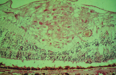
Why cotton wool spots should not be regarded as retinal nerve fibre layer  infarcts | British Journal of Ophthalmology
Why cotton wool spots should not be regarded as retinal nerve fibre layer infarcts | British Journal of Ophthalmology

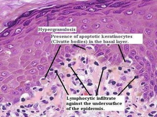
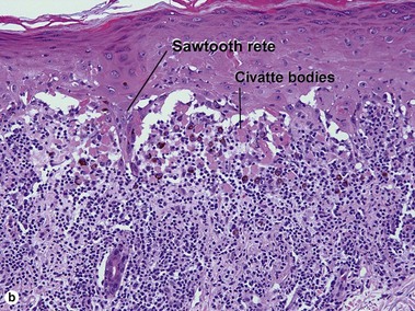
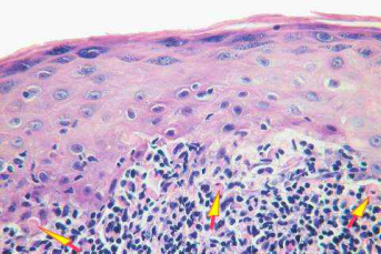
American Society of Dermpath on Twitter: "MT @phmckee1948 Bona fide #Lichen  #Planus. Sawtoothing of rete. Hypergranulosis. Cytoid bodies. Band like  lymphocytic infiltrate. #dermpath https://t.co/dtYXwSeV34" / Twitter
American Society of Dermpath on Twitter: "MT @phmckee1948 Bona fide #Lichen #Planus. Sawtoothing of rete. Hypergranulosis. Cytoid bodies. Band like lymphocytic infiltrate. #dermpath https://t.co/dtYXwSeV34" / Twitter

Rayos, K.- Rodas, F..  21 year old student  CC: Loss of vision OS and eye  aches associated with movement.  PMH: Similar episode in the OD three  years. - ppt download
Rayos, K.- Rodas, F..  21 year old student  CC: Loss of vision OS and eye aches associated with movement.  PMH: Similar episode in the OD three years. - ppt download

Cytoid bodies in cutaneous direct immunofluorescence examination - Wu -  2007 - Journal of Cutaneous Pathology - Wiley Online Library
Cytoid bodies in cutaneous direct immunofluorescence examination - Wu - 2007 - Journal of Cutaneous Pathology - Wiley Online Library

Who Described Civatte Bodies? - Burgdorf - 2014 - Journal of Cutaneous  Pathology - Wiley Online Library
Who Described Civatte Bodies? - Burgdorf - 2014 - Journal of Cutaneous Pathology - Wiley Online Library

Cotton-Wool Spots May Challenge Diagnosis

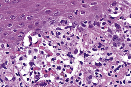
Figure showing skin biopsy of the lesion. Vacuolar alteration of the... |  Download Scientific Diagram
Figure showing skin biopsy of the lesion. Vacuolar alteration of the... | Download Scientific Diagram

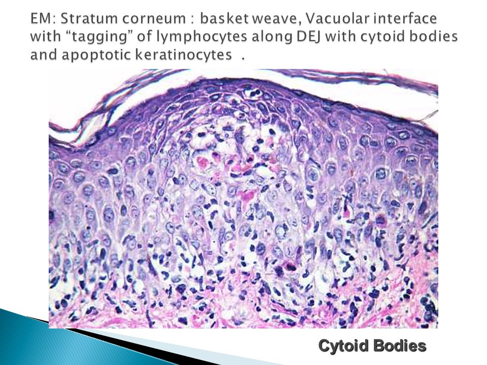
Interface dermatitis | Plastic Surgery Key

Microscopic Description -- Case 68

Civatte bodies: A diagnostic clue Pranay T, Kumar AS, Seema C - Indian J  Dermatol
Civatte bodies: A diagnostic clue Pranay T, Kumar AS, Seema C - Indian J Dermatol

Jerad Gardner, MD on Twitter: "@DrAldehyde @BinXu16 @smlungpathguy  @kriyer68 @Sara_Jiang @DrFNA @bansar_bansaria @VijayPatho More #dermpath  bodies. Civatte/cytoid bodies aka dying/apoptotic/necrotic/dyskeratotic  keratinocytes. Lichen planus here ...
Jerad Gardner, MD on Twitter: "@DrAldehyde @BinXu16 @smlungpathguy @kriyer68 @Sara_Jiang @DrFNA @bansar_bansaria @VijayPatho More #dermpath bodies. Civatte/cytoid bodies aka dying/apoptotic/necrotic/dyskeratotic keratinocytes. Lichen planus here ...

Bodies C-E - Pathological and non-pathological bodies